In August 2020, scientists described one mechanism by which cells â specifically those of a slime mold and mouse pancreatic cancer cells â efficiently navigate through a body and identify the best routes through complex mazes: they generate gradients after breaking down diffused chemoattractants, allowing them to sense upcoming maze junctions prior to reaching them, including around corners. [30] [31] [32] Multicellularity
Monolayer cultures occur when the bottom of the culture jar is completely covered by a continuous layer of cells, typically one cell thick. Monolayers form the majority of continuous cell lines. Due to their single layer structure, these cells may be readily transferred on a cover slip for examination under a microscope. Cells in suspension
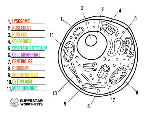
Cellular Architectures
The cell is life's fundamental functional and structural unit. Numerous creatures, such as amoeba and bacteria, are single-celled. The remainder are multicellular, consisting of billions of trillions of cells that comprise an animal's body. A protective membrane surrounds each cell. The cell nucleus is located in the cell's core and holds the genetic information (DNA). Apart from the nucleus, the cell contains several microscopic structures such as ribosomes, mitochondria, and lysosomes, each of which performs one or more critical roles that enable life to exist.
Originally devised to promote clonal expansion of Chinese hamster ovary (CHO) cells, Hamâs Nutrient Mixtures (ATCC CCL-61). As with EMEM, various variations to the original formulation have occurred, including Ham's F-12 medium, a more complicated formulation than F-10 that is ideal for serum-free propagation. Kaighn's version of Hams F-12 (Hams F-12K) was developed to promote primary cell proliferation and differentiation in the presence or absence of serum. F-12K has more amino acids, pyruvate, biotin, calcium, magnesium, putrescine, and phenol red than F-12. ATCC Hamâs F-12K (ATCC 30-2004) has a lower sodium bicarbonate content (1,500 mg/L) and is recommended for use with 5% CO 2.
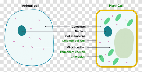
Plant And Animal Cell Picture With Labels
Chloroplasts are chlorophyll-containing organelles that are required for photosynthesis. Photosynthesis is the process through which plants get energy from the sun. Autotrophs are plants that make their own sustenance from sunshine. Animals and other heterotrophs thrive only on organic stuff. Chloroplasts contain their own DNA and are remarkably similar to prokaryotic bacteria; scientists think that chloroplasts were formerly prokaryotic bacteria living within algae 1.5 billion years ago. This kind of contact is referred to as an endosymbiotic relationship. Prokaryotes evolved into chloroplasts inside eukaryotic cells throughout time, and these cells gave birth to several kinds of algae and, eventually, plants.
Membrane of the Cell: This membrane is present in both plant and animal cells. Also known as plasma membrane or plasmalemma, it is the outermost covering of an animal cell, whereas in a plant cell, it is located next to the cell wall. This membrane's primary duties are to form the cell, protect the organelles, and carry nutrients from inside the cell to the outside and vice versa. The nucleus is the cell's control center. It is a spherical-shaped organelle that is found in both plant and animal cells. It contains the chromosomes, which comprise an organism's genetic material. Additionally, protein synthesis starts in this organelle and is finished in the cell cytoplasm. The nucleus contains around 10% of the cell's volume.
What Is the Distinction Between Animal and Plant Cells?
Plant and animal cells are both classified as eukaryotic cells and have a same general structure. However, these two kinds of cells are not identical. The primary distinction between plant and animal cells is that plant cells have a cell wall, while animal cells have not. The form of plant and animal cells is another distinction. Animal cells are amorphous, but plant cells have a distinct rectangular form. Additionally, when it comes to vacuoles, there is a distinction between plant and animal cells. That example, whereas plant cells have a single big vacuole, animal cells contain several tiny vacuoles.
Utilize one of the landscape poster styles to have your pupils identify a plant and animal cell (small or large). Students will design a cell diagram that includes all of the organelles found in plant and animal cells. The cell diagrams are simply colorable, enabling pupils to immediately distinguish between the many components of a plant or animal cell. By grouping them together on a single board, students can rapidly grasp the distinctions between the cells, such as the organelles that plant cells possess but animal cells do not.
Animal Cell Picture Without Labels
Consider how a single-celled creature has the structures essential for eating, growing, and reproducing. Cells are the fundamental building blocks of life. Inc. Encyclopaedia Britannica View all videos associated with this article This article focuses on animal cells, with some discussion of plant-specific energy-synthesizing mechanisms and extracellular components. (See photosynthesis for a thorough description of the biology of plant cells.) For a comprehensive discussion of the genetic processes that occur inside the cell nucleus, see heredity.)
Karyokinesis, alternatively called mitosis, is a succession of stages that culminate in the division of the cell nucleusâprophase, prometaphase, metaphase, anaphase, and telophase (Figure 2). Karyokinesis is another term for mitosis. The Artistic Connection Which of the following statements is right about the sequence of events during mitosis? At the metaphase plate, sister chromatids align. The kinetochore is connected to the mitotic spindle during mitosis. The nucleus reorganizes and the cell division occurs. Cohesin proteins degrade, resulting in the separation of sister chromatids. The kinetochore is connected to the mitotic spindle during mitosis. Cohesin proteins degrade, resulting in the separation of sister chromatids. At the metaphase plate, sister chromatids align. The nucleus reorganizes and the cell division occurs. The kinetochore is cohesin-protein-coated. At the metaphase plate, sister chromatids align. The kinetochore disintegrates, resulting in the separation of sister chromatids. The nucleus reorganizes and the cell division occurs. The kinetochore is connected to the mitotic spindle during mitosis. At the metaphase plate, sister chromatids align. Cohesin proteins degrade, resulting in the separation of sister chromatids. The nucleus reorganizes and the cell division occurs.
Rat oviduct cilia and mucous cells. Getty Images / Micro Discovery Cells are the basic living units in the hierarchical framework of life. Animal creatures may be billions of cells in size. There are hundreds of distinct cell kinds in the human body. These cells come in a variety of forms and sizes, and their architecture is optimized for their purpose. For instance, brain cells, or neurons, in the body have a radically different form and function than red blood cells. Nerve cells are responsible for the transmission of electrical impulses throughout the nervous system. They are elongated and thin, with protruding projections that connect to other nerve cells in order to conduct and transmit nerve impulses. Red blood cells' primary function is to carry oxygen to bodily cells. Their compact, flexible disc form allows them to go through microscopic blood channels, delivering oxygen to organs and tissues.
Cellular Architectures
The cell is life's fundamental functional and structural unit. Numerous creatures, such as amoeba and bacteria, are single-celled. The remainder are multicellular, consisting of billions of trillions of cells that comprise an animal's body. A protective membrane surrounds each cell. The cell nucleus is located in the cell's core and holds the genetic information (DNA). Apart from the nucleus, the cell contains several microscopic structures such as ribosomes, mitochondria, and lysosomes, each of which performs one or more critical roles that enable life to exist.
Animal Cell Picture No Labels
Bacterial infections, such as Mycoplasma, and fungal infections often develop in cell culture, posing an identification and elimination challenge. Thus, all cell culture experiments are conducted in a sterile setting using aseptic procedures. Work should be carried out in a laminar flow environment with a continual unidirectional flow of HEPA-filtered air passing over the work area. All materials, solutions, and the whole environment should be free of contamination. Cryopreservation
Endosomes and Endocytosis - Endosomes are membrane-bound vesicles created by a complex set of processes generally referred to as endocytosis. They are present in the cytoplasm of almost every animal cell. Endocytosis is fundamentally the inverse of exocytosis or cellular secretion. It is a process in which the plasma membrane of a cell invaginates (folds inward) to envelop macromolecules or other materials diffusing through the extracellular fluid. Golgi Apparatus - The Golgi apparatus serves as the cell's distribution and shipping department. It alters proteins and lipids synthesized in the endoplasmic reticulum in preparation for their export to the extracellular space.
" " All living things, plant and animal alike, are composed of remarkably identical tiny building pieces known as cells, which serve as the fundamental structural and functional components of life. Getty Images/HowStuffWorks jack0m From the exterior, plants seem to be very distinct from mammals. For example, plants are unable to move and catch food in the same manner that humans do, they emit oxygen instead of carbon dioxide, and they lack the sense organs that enable us to avoid a fire or smell out and hunt down a prospective meal. However, plants and animals are more alike than they seem on the surface. Indeed, a plant cell and an animal cell may seem so similar under a microscope that in certain circumstances, you'd really have to know what you're looking at to tell them apart.
LysosomesIn animal cells, lysosomes function as the cell's âgarbage disposal system.â Within the lysosomes, digestive enzymes help in the digestion of proteins, polysaccharides, lipids, nucleic acids, and even worn-out organelles. In single-celled eukaryotes, lysosomes are required for food digestion and organelle recycling. These enzymes are more acidic in nature than those found in the cytoplasm. Numerous processes that occur in the cytoplasm cannot proceed at a low pH, emphasizing the need of compartmentalizing the eukaryotic cell into organelles.